

A new rapid protein transport system has been discovered.
Scientists have discovered a previously unknown intracellular system that speeds up the delivery of proteins to the leading edge of the cell. This finding changes our understanding of the mechanisms behind cell movement, cancer spread, and wound healing, and could also lead to new strategies for combating tumors.
Cursus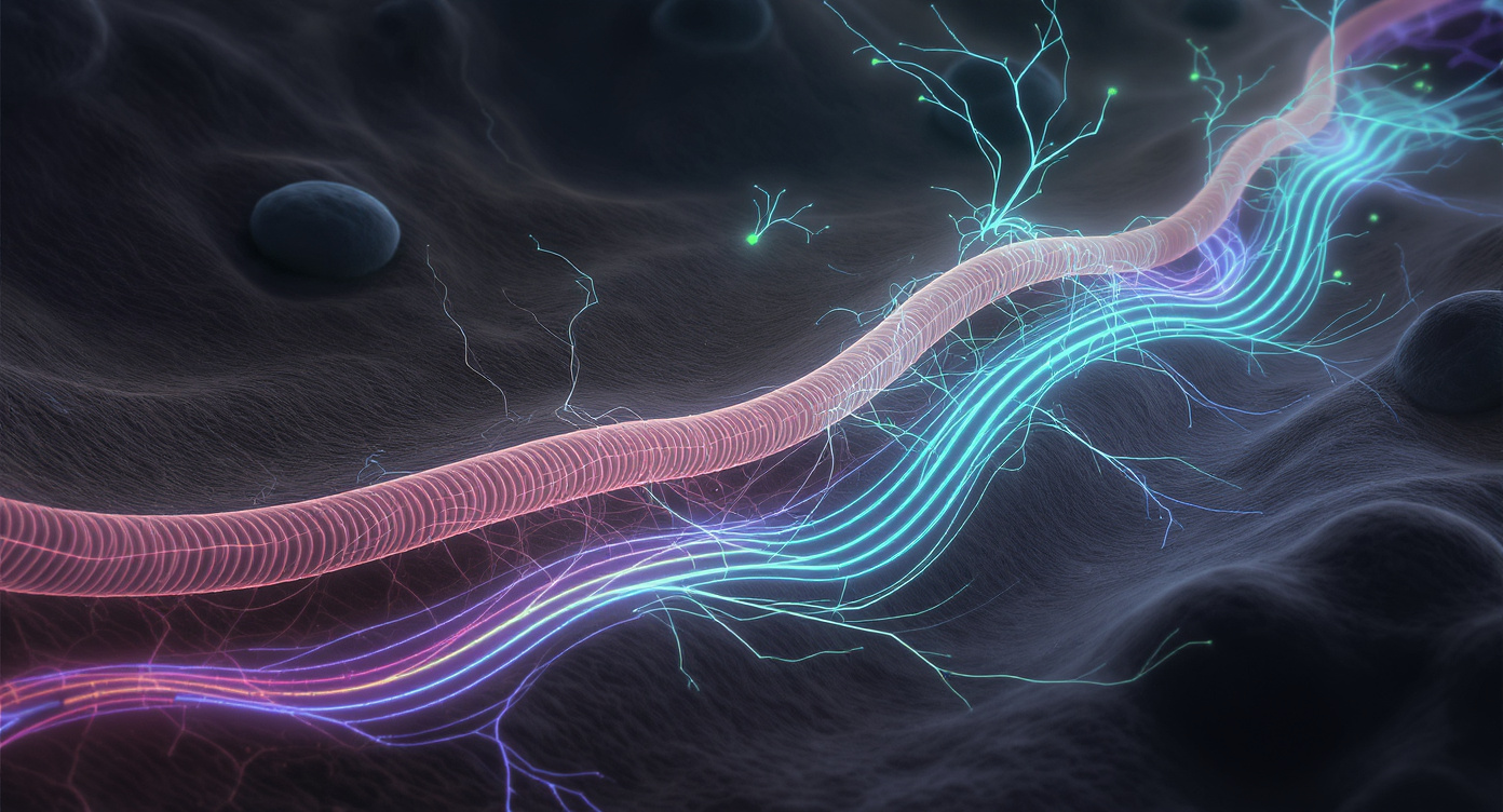
Researchers from the Oregon Health & Science University have discovered a previously unknown intracellular system that enables rapid transport of proteins to the leading edge of the cell. This finding reshapes our understanding of the mechanisms behind cell movement, cancer spread, and wound healing.
A New System for Intracellular Transport
It was previously believed that protein transport within cells occurred mainly through random diffusion, where molecules move chaotically until they reach their destination. However, a new study published in Nature Communications has shown that cells use directed fluid flows to actively move proteins such as actin to the leading edge, where growth, movement, and tissue repair take place.
Experimental Observations
During experiments using laser microscopy, scientists temporarily made proteins invisible in a specific region of the cell to track their movement. As a result, they observed an additional dark band at the cell’s leading edge—in the area that extends during movement. Further analysis revealed that this band represents a wave of soluble actin rapidly advancing forward, indicating a different protein delivery mechanism than random diffusion.
The Mechanism of Directed Flows
Using specialized visualization techniques, researchers established that cells generate directed fluid flows that transport actin and other proteins to the leading edge much faster than diffusion alone would allow. These flows are non-specific and can carry several types of proteins at once, ensuring efficient support for the movement of cellular protrusions, attachment, and rapid shape changes necessary for cell motility, immune responses, and tissue repair.
Structural Features
The study showed that these flows arise in a specialized region at the cell’s leading edge, separated from the rest of the cell by a condensate of actin and myosin, which acts as a physical barrier and directs proteins toward the advancing edge.
Technologies and Methods
To observe internal flows, the team developed a modified version of the fluorescence microscopy method, activating fluorescent molecules at a single point and tracking their movement. One of the key experiments was named FLOP (Fluorescence Leaving the Original Point). The research also utilized advanced visualization tools, including the iPALM technique, which allows for distinguishing structures at the nanometer scale.
Significance for Medicine and Biology
This discovery may explain the high aggressiveness of certain cancer cells that possess this rapid protein delivery mechanism to the leading edge. Understanding the differences in how this system is used by cancerous versus normal cells could help develop new strategies to slow or stop tumor spread.
An Interdisciplinary Approach
The research involved experts in engineering, physics, microscopy, cell biology, as well as specialists in fluorescence spectroscopy and 3D visualization from the Janelia Research Campus.
Application Prospects
The discovered system is described as a functional compartment not bounded by a membrane, yet playing a crucial role in organizing cell behavior. This breakthrough is expected to influence cancer research, drug delivery methods, tissue regeneration, and synthetic biology.

k6 comment VU-147