

Magnetic Particle Tomography: A New Era in Vascular Diagnostics
For the first time, magnetic particle tomography has been tested on a human—a technology that enables safe monitoring of blood flow without the use of X-ray radiation. This method could become a new tool for vascular medicine and expand the possibilities of real-time diagnostics.
Salus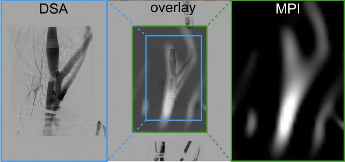
Study: First Demonstration of Magnetic Particle Imaging in Humans
A New Method for Medical Imaging
For the first time, magnetic particle imaging (MPI) has been tested on a human subject. This technology enables real-time tracking of blood flow through the veins of the arm without the use of X-ray radiation, potentially offering a safer alternative to traditional vascular angiography.
The Evolution of Imaging Techniques
Since the discovery of X-rays in 1895, medical imaging has become a crucial diagnostic tool. Modern methods include computed tomography (CT), magnetic resonance imaging (MRI), ultrasound, and positron emission tomography (PET), each with its own advantages and limitations.
CT and X-ray imaging involve ionizing radiation, and the contrast agents used in angiography can be problematic for patients with kidney disease. As a result, researchers are seeking safer ways to observe blood flow and assess vascular health.
How Magnetic Particle Imaging Works
Magnetic particle imaging, introduced in 2005, differs from MRI in that it does not directly visualize tissues. Instead, iron oxide nanoparticles are injected into the bloodstream, and a specialized scanner generates a varying magnetic field to detect the response of these particles. Since human tissues produce virtually no such signal, the resulting images have minimal background noise, allowing for rapid and precise tracking of particle distribution, such as blood movement through vessels.
Development and Testing of the Technology
Over the past two decades, the technology has been refined in laboratory settings and animal models. The main challenge was to develop sufficiently large scanners and ensure safe magnetic field parameters for clinical use.
A research team from the University of Würzburg developed an interventional MPI scanner that can be placed directly in an angiography suite. In the first-ever human experiment, a clinically approved iron nanoparticle-based agent diluted in saline was administered to a volunteer. The MPI scanner was then used to monitor the distribution of the particles through the veins of the arm. For comparison, a traditional X-ray angiography was also performed.
Results and Future Prospects
The new method allowed visualization of the same major veins as the X-ray procedure, including both superficial and deep vessels, branches, venous valves, and the direction of blood flow. The system operated in real time at a rate of about two frames per second. Researchers were also able to observe how the magnetic contrast gradually left the vessels, enabling dynamic monitoring of blood circulation.
No side effects were observed during the procedure, and both the magnetic field parameters and energy absorption levels were well below established medical safety limits.
Limitations and Further Development
Currently, the technology is in its early stages: the prototype scanner has a relatively small field of view and limited spatial resolution. Further improvements in hardware are expected to address these challenges.
Significance for Medicine
Magnetic particle imaging could become a valuable tool in vascular medicine, especially for endovascular surgery. If successfully developed, this technology could significantly enhance existing medical imaging methods and open new possibilities for real-time observation of blood flow, cellular processes, and vascular function.