

Unexpected Behavior of the T-Cell Receptor Revealed by Scientists
A new study has shown that the T-cell receptor remains closed until it encounters an antigen, rather than always being open as previously believed. This discovery could help improve immunotherapy and vaccine development.
Cursus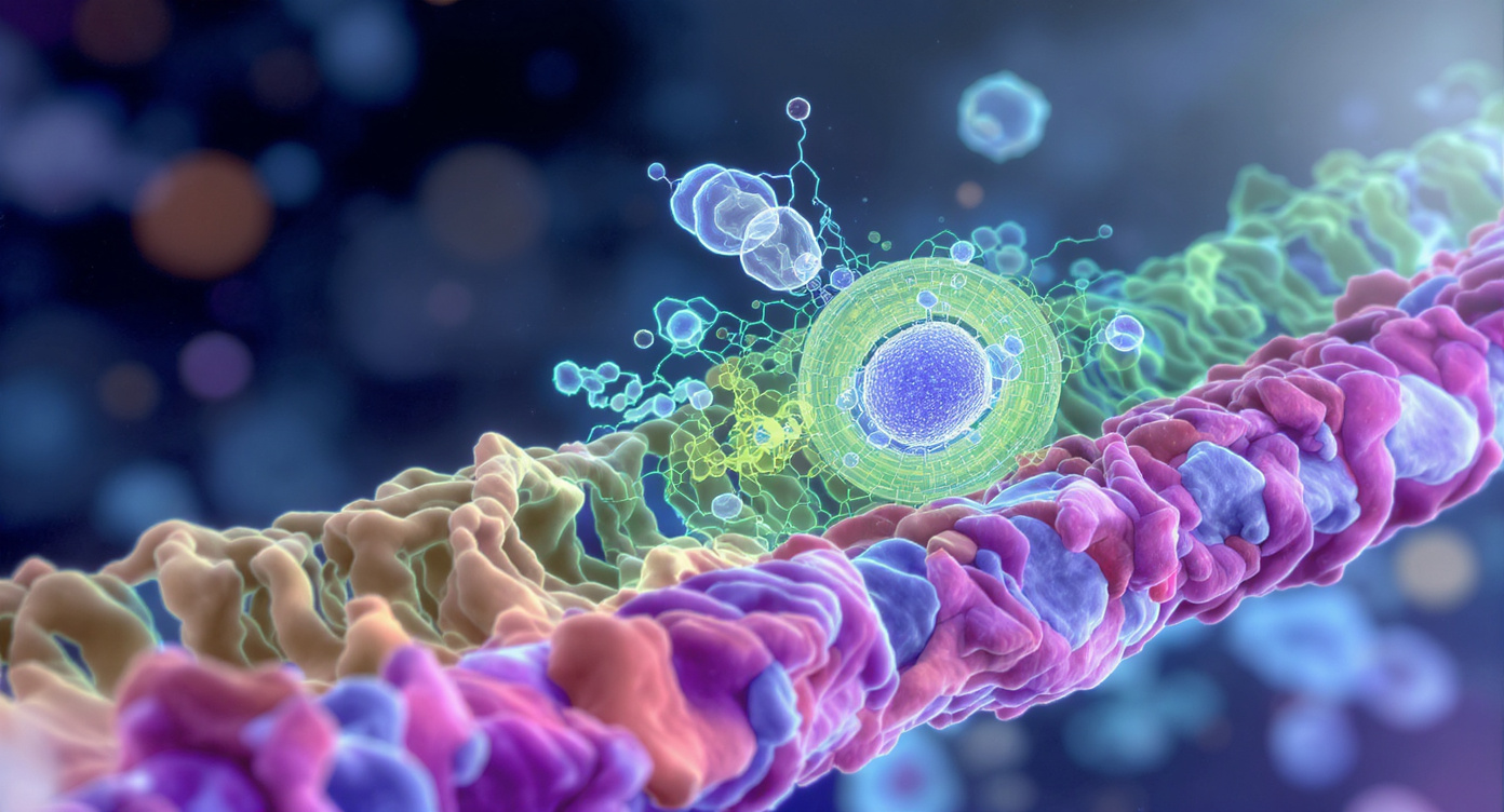
Over the past decade, T-cell immunotherapy has emerged as one of the most promising approaches for cancer treatment. These methods are based on training the patient’s immune system to recognize and destroy dangerous cells. Despite significant progress, scientists still do not fully understand how these therapies work at the molecular level. This lack of knowledge slows the development of new treatments, especially since T-cell methods are only effective for certain types of cancer, and the reasons for their ineffectiveness in others remain unclear. A deeper understanding of these mechanisms could help make these therapies accessible to more patients.
New Discoveries in T-Cell Receptor Structure
Researchers at Rockefeller University have uncovered important details about the T-cell receptor (TCR)—a protein complex embedded in the cell membrane that plays a key role in T-cell therapies. Using cryo-electron microscopy (cryo-EM), scientists from the molecular electron microscopy lab studied the TCR in conditions that closely mimic its natural environment. They found that the TCR behaves like a “spring in a box”: it remains compact until it encounters an antigen or another suspicious particle, at which point it rapidly unfolds. This behavior contradicts previous studies, which suggested the receptor was always in an open state. These new findings could help improve and expand the use of T-cell immunotherapies.
The Importance of the Membrane Environment for Receptor Function
The Walz laboratory specializes in creating detailed images of macromolecular complexes, especially proteins located in cell membranes that connect the cell to its environment. The TCR is one such complex, made up of several proteins, allowing T-cells to recognize antigens presented by HLA complexes on other cells. This process is fundamental to T-cell therapies, which mobilize the immune system against cancer.
Although individual parts of the TCR have been known for a long time, the initial stages of its activation remained unclear. This is particularly important for patients with sarcomas, who often do not benefit from T-cell immunotherapies. Understanding these processes will help clarify how information is transmitted from outside the cell, where antigens are presented by HLA, to the inside of the cell, where signaling is triggered and the T-cell is activated.
Technological Advances in TCR Research
The Walz team is known for creating artificial membrane environments that closely resemble natural conditions for membrane proteins. In this study, scientists placed the receptor in a nanodisc—a tiny disc-shaped section of membrane held together in solution by a protein scaffold around the edge. Assembling the full receptor was a complex task, and correctly integrating all eight proteins into the nanodisc required significant effort.
Previously, structural studies of the TCR were conducted using detergents, which often remove the surrounding membrane. For the first time, the receptor complex was reconstituted in a membrane environment for detailed study.
Unexpected Results and Their Significance
After embedding the TCR in the nanodisc, researchers used cryo-EM to visualize it. The images showed that the receptor remains closed and compact in its inactive state. When it encounters an antigen molecule, the structure opens and extends outward, resembling a broad gesture.
This result surprised the team, as it was previously believed that the complex was open even at rest and did not change shape upon binding to antigens. However, it turned out that the receptor does indeed change conformation, unfolding like a “spring in a box.”
The researchers believe their discovery was made possible by two factors: first, they recreated the TCR’s membrane environment using the correct lipid mixture; second, they embedded the receptor in the membrane with nanodiscs before performing cryo-EM. It turned out that an intact membrane keeps the receptor closed until activation. In previous studies, detergents may have removed this restriction, allowing the receptor to open prematurely.
Using a lipid mixture that closely resembles the T-cell membrane proved to be critically important: if a model lipid environment had been used, the closed state of the receptor would not have been detected.
Implications for Therapy and Vaccines
The team is confident that their discoveries will help improve T-cell receptor-based treatment methods. For example, it may become possible to adjust receptor sensitivity by changing their activation threshold, which is especially important for treating rare sarcomas. Additionally, the data obtained could be useful for vaccine development: specialists can now use new structural information to study interactions between different antigens presented by HLA and T-cell receptors, potentially optimizing their functions.