

Cells transport proteins using microscopic pumps.
A new study has shown that soluble proteins inside migrating cells move thanks to directed fluid flows generated by contracting protein structures. This physical mechanism enables cells to rapidly deliver molecules to specific areas without relying on specialized chemical pathways.
Cursus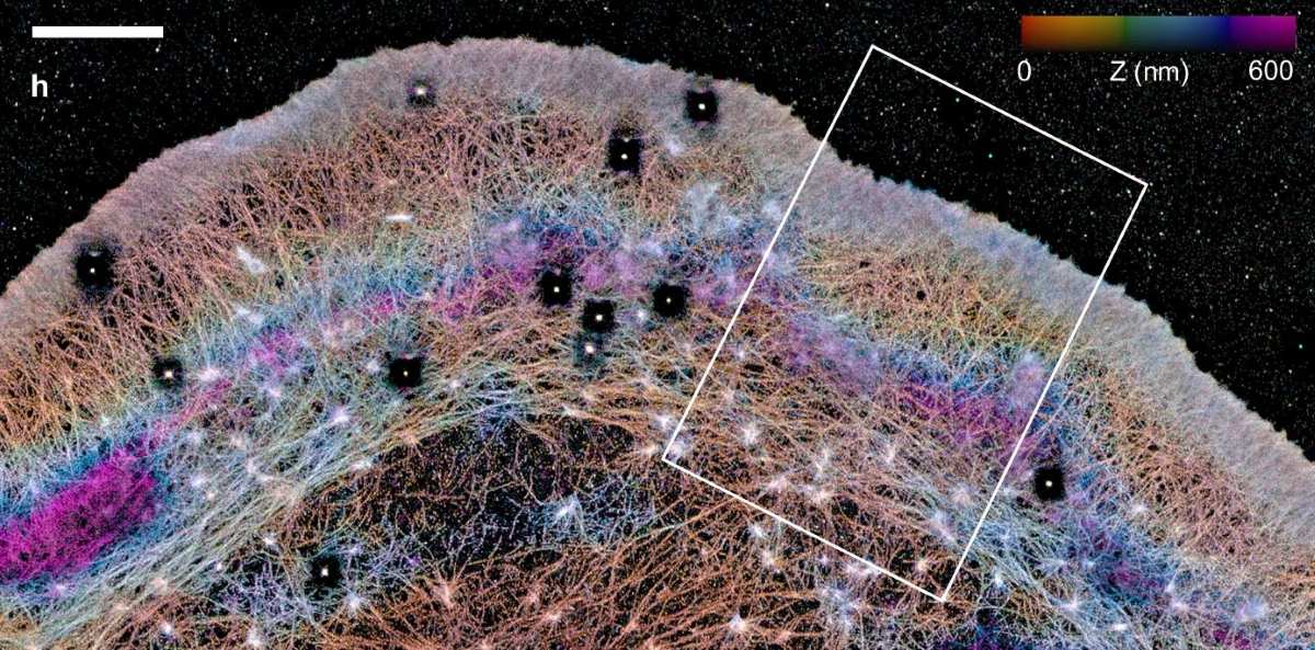
Soluble proteins inside migrating cells do not move randomly, but are transported by directed flows of liquid. These flows are generated by a special non-membranous barrier made up of contracting molecules, which acts like a microscopic pump, delivering dissolved substances to the leading edge of the cell.
Mechanism of Soluble Molecule Transport
During cell movement, it is essential to continuously supply the leading edge with new molecules needed to build the cell’s cytoskeleton and membrane. Large organelles and vesicles travel along microtubules with the help of motor proteins, but soluble molecules lack such “engines.” Previously, it was believed that these proteins spread through the cytoplasm by random diffusion, but mathematical models have shown that this process is too slow and cannot provide the necessary speed for cell movement.
Experimental Observations
In a recent study, researchers developed the FLOP (Fluorescence Leaving the Original Point) microscopy method, which allows tracking the movement of dissolved substances within a cell. Mouse neuroblastoma cells and fibroblasts were engineered to express photoactivatable proteins that begin to fluoresce under ultraviolet light. During the experiment, a laser illuminated a single point inside the cell, and the spread of the glowing spot was recorded.
The results showed that soluble molecules move toward the cell’s leading edge in a directed flow at a speed of about 3.6 micrometers per second, which is nearly 50 times faster than the movement of cytoskeletal filaments in the opposite direction. These flows have been termed “cytoplasmic trade winds,” by analogy with the steady winds between the tropics.
Testing the Mechanical Nature of the Process
To test the mechanism of the transport system, the function of the motor protein myosin II was blocked using the inhibitor blebbistatin. After this, the flows stopped completely, and molecules began to spread slowly and symmetrically, as in ordinary diffusion.
Three-dimensional visualization revealed that the front part of the cell (the lamella) is separated from the rest of the cell body by a dense, arc-shaped wall of actin and myosin molecules. This structure functions as a non-membranous protein barrier that constantly contracts, pushing the intracellular fluid forward. The barrier acts like a semi-permeable dam: it allows liquid to pass toward the cell edge but hinders the backward flow of proteins.
Universality of the Mechanism
The study revealed the non-specific nature of this “pump”: unlike molecular motors, which recognize only certain cargos, intracellular flow (advection) carries all elements—actin monomers, cell adhesion proteins, and even artificially introduced inert fluorescent dyes. When the protein barrier was locally destroyed by a laser, the flow was disrupted, and the leading edge of the cell at that site immediately stopped and retracted inward.
Significance for Cell Movement
Cells use the laws of hydrodynamics to control their own movement. The formation of dynamic compartments with contracting protein walls allows cytoplasm to be pumped and resources to be concentrated in the necessary areas. This physical mechanism explains how biological systems coordinate the directed transport of molecules without the involvement of specialized chemical pathways or isolating lipid membranes.