

AI has learned to predict the progression of osteoarthritis from images
Researchers from the University of Surrey have developed an AI system capable of predicting what a knee joint X-ray will look like a year in advance, opening up new possibilities for diagnosing and treating osteoarthritis.
Salus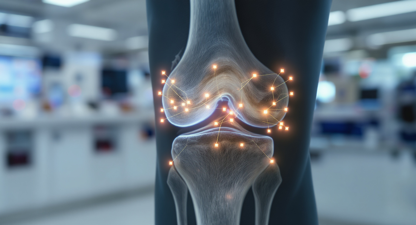
Scientists at the University of Surrey have developed an artificial intelligence system capable of predicting what a patient’s knee X-ray will look like a year into the future. This innovation opens up new possibilities for diagnosing and treating osteoarthritis—a condition that affects more than 500 million people worldwide.
The new AI model was presented at the International Conference on Medical Image Computing and Computer Assisted Intervention (MICCAI 2025). The system generates realistic future X-ray images of joints and provides personalized forecasts for disease progression. This approach gives doctors and patients a clear visual understanding of how osteoarthritis may advance over time.
Osteoarthritis is a degenerative joint disease and is considered the leading cause of disability among older adults. To train the system, researchers used around 50,000 knee X-rays from approximately 5,000 patients—one of the largest datasets of its kind. The technology predicts disease progression about nine times faster than existing methods, while also offering greater accuracy and efficiency.
The core of the development is a generative diffusion model. It creates predictive X-ray images and identifies 16 key points in the joint, highlighting areas where changes are likely to occur. This makes the AI’s work transparent for doctors, showing exactly which parts of the knee the system is analyzing.
The researchers believe this technology could also be applied to other chronic diseases. In the future, similar systems may be able to predict lung damage in smokers or track the development of cardiovascular conditions. The team is currently seeking partners to implement the technology in clinical practice.
Previous AI systems assessed the risk of osteoarthritis progression slowly and only provided numerical indicators without visualization. The new approach allows for rapid creation of clear predictive images and precise identification of problematic joint areas, helping doctors to promptly identify at-risk patients and select personalized treatment plans.