

Expectations are formed in the brain as a distributed network.
A new large-scale study has revealed that expectations and decision-making are not formed in separate centers of the brain, but rather through a distributed network spanning multiple regions. This discovery changes our understanding of how the brain works and highlights its complex, interconnected structure.
Cursus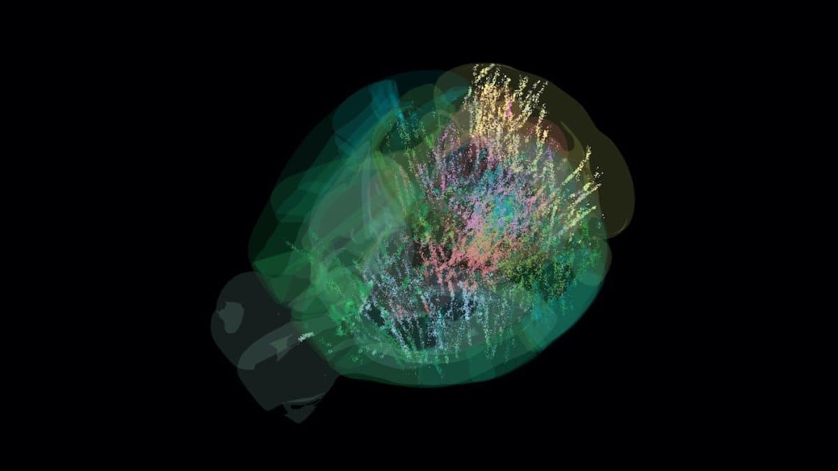
A large-scale study of neural activity in mice has offered a fresh perspective on how the brain makes decisions. The findings reveal that this process does not rely on a single specialized region, but rather on a distributed network spanning nearly the entire brain—from sensory to motor areas.
From Local Centers to Distributed Networks
For many years, neurobiologists have explored complex cognitive functions like decision-making by focusing on small groups of cells in specific brain regions. This approach assumed the existence of narrowly specialized “decision centers.” However, the brain is an extraordinarily complex and interconnected system, where thousands of neurons from hundreds of regions are constantly exchanging information. In recent decades, it has become clear that studying isolated regions does not provide a complete picture.
The Problem of Fragmented Data
The main challenge was that different laboratories studied different parts of the brain, using various tasks and analysis methods. Integrating such fragmented data into a unified model was impossible: even the mouse brain is too large and complex, and the experiments too diverse, for each to illuminate more than a small part of the “neural landscape.”
A New Approach: Standardization and Scale
To address this, the International Brain Laboratory (IBL) was formed, bringing together 22 labs from Europe and the USA. The researchers developed a single, strictly standardized experiment. In it, 139 mice were trained to perform a seemingly simple task: the animal sat in front of a screen and used a small steering wheel to move a striped circle, which appeared on the left or right, to the center of the screen. For correct and quick actions, the mouse received a drop of sweet water as a reward.
The key feature of the task was its probabilistic structure. During blocks of 20–100 trials, the circle appeared on one side 80% of the time and on the other 20%. Then, the probabilities would switch without any signal to the animal. To maximize their reward—especially in trials with zero contrast (when the stimulus was invisible)—the mice had to track this hidden pattern and form an internal expectation, or “prior probability.” This allowed researchers to study how expectations influence future decisions.
Large-Scale Data and New Discoveries
While the mice performed the task, scientists recorded neural activity using hundreds of high-density Neuropixels electrodes. This resulted in a unique dataset: activity from 621,733 neurons across 279 brain regions. Analysis showed that mice did indeed use the probabilistic structure of the task to improve their performance, especially in trials without a visible stimulus—their accuracy reached nearly 59%, significantly above chance.
Information about the prior probability—the animal’s internal expectation—was not encoded in just a few specialized regions, but was distributed throughout the brain. Traces of this signal were found in about 30% of all studied regions, covering all levels of information processing: from early sensory zones (such as the primary visual cortex and thalamus) to associative areas and motor centers.
How the Brain Forms Expectations
This discovery challenges models in which expectations are only considered at the final stages of decision-making. The results support the hypothesis that the brain functions as a vast Bayesian network with a constant, multidirectional flow of information. Further analysis revealed another important detail: mice formed their expectations not based on an ideal mathematical model, but on a simpler heuristic. Their internal expectation relied more on their own recent actions than on the presented stimuli.
In other words, the animal adjusted its strategy based on what it had done in the last five or six trials. Neural activity in the brain precisely reflected this subjective, action-based model, rather than the objective probability of stimulus appearance. Signals related to movement and reward were the most widespread—found in nearly all studied brain regions. The representation of choice was also broadly distributed, while the encoding of the visual stimulus itself was limited to classic visual pathways.
Limitations and Future Directions
Despite the scale of the work and its “revolutionary” conclusions, the authors note several limitations related to the complexity of the processes studied. First, signals associated with reward (positive feedback) are difficult to distinguish from neural activity responsible for accompanying movements—such as licking. Second, even with such detailed analysis, much of the recorded neural activity remains unexplained within the context of the task. This may indicate that the brain is constantly engaged in processing internal states or responding to unmonitored movements unrelated to the task.
Thus, even with the creation of the most detailed neural map to date—covering the entire brain—this is only the first step toward understanding how unified, coherent behavior emerges from the distributed activity of billions of neurons, even within what seems to be a thoroughly studied model of the mouse brain.