

Mini-brains may help select treatments for Alzheimer's disease
Scientists have demonstrated that miniature brain models grown from the cells of patients with Alzheimer's disease can help predict individual responses to treatment and identify new disease biomarkers. This approach paves the way for more precise and personalized therapies.
Cursus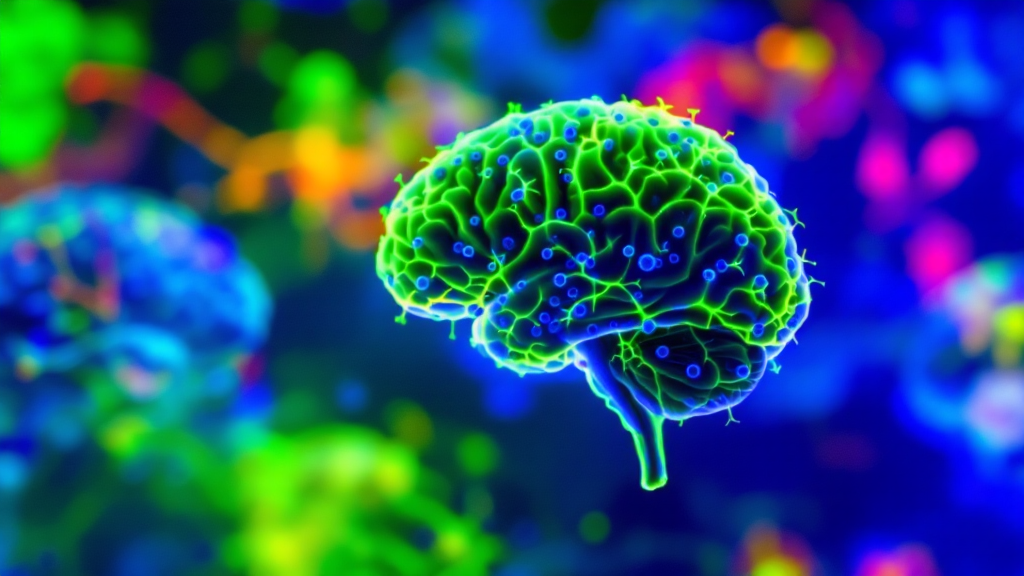
Exploring Miniature Brain Models for Alzheimer's Disease Research
Fluorescent imaging of a brain organoid
Fluorescent microscopy of a brain organoid reveals a neuroepithelial structure, with neurons stained green (TUJ1) and cell nuclei stained blue (DAPI).
Using Organoids to Predict Treatment Response
During the study, small clusters of brain tissue were grown from cells of patients with Alzheimer's disease. These mini-brain models were used to assess individual responses to medications prescribed for managing psychiatric symptoms associated with the disease. The research focused on lab-grown brain organoids. The findings suggest that such models could help develop more precise therapies for different groups of Alzheimer's patients. In the United States, more than 7 million people are affected by this condition.
The study also found that organoids release extracellular vesicles—microscopic particles that carry cellular information. These vesicles are being considered as potential biomarkers for diagnosing Alzheimer's disease and determining its stage of progression.
A Personalized Approach to Therapy
Mini-brain models can support a personalized approach to treatment. Currently, there are no drugs that completely cure Alzheimer's disease, but selective serotonin reuptake inhibitors (SSRIs) are often prescribed to manage symptoms such as anxiety, depression, and agitation. These symptoms are common among patients, but individual responses to medications can vary significantly.
Creation and Analysis of Organoids
The research was conducted on miniature models of the hindbrain—the region responsible for breathing, sleep, and heart rhythm. Scientists aimed to determine whether these models could detect molecular markers of the effectiveness of escitalopram oxalate, a drug used to reduce Alzheimer's symptoms.
To create the organoids, blood samples from both Alzheimer's patients and healthy individuals were used. The cells were reprogrammed into induced pluripotent stem cells, which can develop into any cell type in the body. From these cells, hindbrain organoids were formed, containing specialized neurons that produce serotonin. The cells self-organized into small clusters resembling the hindbrain. Hundreds of organoids were included in the study, representing both patients and healthy participants.
Biological Features and Treatment Response
Organoids derived from Alzheimer's patient cells exhibited several characteristic molecular features of the disease. Compared to organoids from healthy individuals, they showed differences in proteins involved in cell-to-cell communication, inflammation, and other disease-related pathways.
After treatment with escitalopram, some organoid samples showed increased levels of proteins associated with serotonin signaling and intercellular communication—these are the pathways targeted by antidepressants. However, other organoids showed little to no molecular response to the treatment.
Extracellular Vesicles as Potential Biomarkers
The study also investigated whether extracellular vesicles released by organoids could serve as biomarkers for Alzheimer's disease or help assess tissue response to treatment. Proteins inside vesicles from both patient-derived and control organoids were analyzed before and after escitalopram treatment.
The vesicles contained proteins involved in key brain processes, including neuron-to-neuron communication, memory, and neurotransmitter release. Organoids from Alzheimer's patients showed significant changes in several disease-related proteins. Levels of RAB3A, NSF, and ATCAY proteins were lower in Alzheimer's organoids, which is important for normal signal transmission between brain cells.
After escitalopram treatment, some samples showed increased levels of certain proteins, especially those linked to serotonin signaling and synaptic pathways. Some organoids demonstrated strong molecular responses, while others changed very little. This diversity suggests that extracellular vesicles could help identify patients most likely to benefit from specific treatments.
Future Prospects for Organoid Models
There are plans to develop more complex organoids that include immune cells and vascular structures to better mimic human brain tissue. In the future, extracellular vesicles may be used as a "liquid biopsy" for diagnosing Alzheimer's disease, determining its stage, and identifying subtypes in individual patients. This current research is seen as a first step in that direction.